3.1. Process of Seeing and Common Eye Disorders in India
The eye is the apparatus for seeing a general knowledge of the eye & how it works is useful in helping to understand impaired vision.
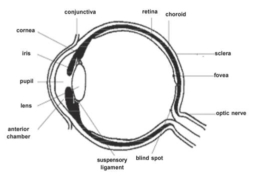
The Eye: The eye & brain work together to form images & the things we see. We have two eyeballs. Each eyeball is about 2.5cms. in diameter and is lodged within the socket called orbit between the orbital wall & the eyeball.
Eyelids: These are movable folds of skin which protect the eye from injury and excessive light. The eyebrows and eyelashes also participate in the protective role.
The conjunctiva: It is a continuous sheet of tissue. To allow free movements of the eyeball, the conjunctiva is carried above & below into folds.
Cornea: Cornea is a crystal clear structure in front surface of the eye. It carries the light rays to the retina in the back of the eye.
Sclera: Sclera is the white of the eye which joins the cornea at the limbos. It is tough and helps to maintain the shape of the eye and supports the delicate structures within the eye.
Anterior Chamber: This is behind the cornea and in front of the iris and lens. A clear aqueous fluid secreted by the processes of the ciliary body flows constant through the pupil into the anterior chamber & maintains constant pressure within the eyeball.
Iris: The black disc behind the cornea is called iris. It is an extremely delicate, constantly moving diaphragm with a circular opening in middle. It contains pigments, which give colour to the eyes. These pigments control the amount of light entering retina.
Cilliary Body: Cilliary body is the portion of the veal tract between the iris and the choroids.
Choroid: This is vascular, intermediate coat which furnishes nourishment to the other parts of the eyeball.
Crystalline lens: This is transparent, colourless body suspended in the eyeball.
Vitreous humour: Transparent, colourless mass of soft, gelatinous material filling the eyeball behind the lens.
Retina: This is the innermost coat of the eye, formed of light sensitive nerve element. The most specialised part of the retina is ‘macula’ which lies just lateral to the optic disc. It provides acute central vision used in reading or in threading a needle.
How We See: The eye functions like a camera. Vision is a complex function. The act of seeing requires light to see by and the brain to interpret what is seen. The light rays reflected from an object in a person’s field of vision fall on the eyes.
The rays pass through the cornea, aqueous humour, and through pupil which dilates or contracts to control light in accordance to the brightness of the object. The rays then pass through crystal lineless and the rays of light are focussed on the retina. The process of focusing is called accommodation. The cornea and the lens combine to bend the light rays as they pass through. The rays pass through the vitreous body and penetrate on retina, where they set up a photo-chemical response in the outermost layers, stimulating the rods & cones.
The impulse is picked by retinal nerve fibres and passes along the optic nerve to the brain where an upside down image is formed. Based on experience, the inverted image is psychologically transposed.
Common Eye Disorders
Refractive errors are the most frequent eye problems. Refractive errors include myopia (near-sightedness), hyperopia (farsightedness), astigmatism (distorted vision at all distances), and presbyopia that occurs between age 40–50 years (loss of the ability to focus up close, inability to read letters of the phone book, need to hold newspaper farther away to see clearly) can be corrected by eyeglasses, contact lenses, or in some cases surgery.
Retinitis Pigmentosa is a genetic or inherited condition. Initially it manifests as night blindness. As the disease progresses there may be a tunnelling of vision with loss of peripheral vision followed by complete blindness.
Macular degeneration, often called age-related macular degeneration (AMD), is an eye disorder associated with aging and results in damaging sharp and central vision. Central vision is needed for seeing objects clearly and for common daily tasks such as reading and driving. AMD affects the macula, the central part the retina that allows the eye to see fine details.
Cataract is a clouding of the eye’s lens and is the leading cause of blindness worldwide, and the leading cause of vision loss in the United States. Cataracts can occur at any age because of a variety of causes, and can be present at birth.
Diabetic retinopathy (DR) is a common complication of diabetes. It is characterized by progressive damage to the blood vessels of the retina, the light-sensitive tissue at the back of the eye that is necessary for good vision. DR progresses through four stages, mild nonproliferative retinopathy (microaneurysms), moderate nonproliferative retinopathy (blockage in some retinal vessels), severe nonproliferative retinopathy (more vessels are blocked leading to deprived retina from blood supply leading to growing new blood vessels), and proliferative retinopathy (most advanced stage). Diabetic retinopathy usually affects both eyes.
Glaucoma is a group of diseases that can damage the eye’s optic nerve and result in vision loss and blindness. Glaucoma occurs when the normal fluid pressure inside the eyes slowly rises. However, recent findings now show that glaucoma can occur with normal eye pressure. With early treatment, you can often protect your eyes against serious vision loss.
Amblyopia, also referred to as “lazy eye,” is the most common cause of vision impairment in children. Amblyopia is the medical term used when the vision in one of the eyes is reduced because the eye and the brain are not working together properly. The eye itself looks normal, but it is not being used normally because the brain is favoring the other eye. Conditions leading to amblyopia include strabismus, an imbalance in the positioning of the two eyes; more nearsighted, farsighted, or astigmatic in one eye than the other eye, and rarely other eye conditions such as cataract.
Strabismus involves an imbalance in the positioning of the two eyes. Strabismus can cause the eyes to cross in (esotropia) or turn out (exotropia). Strabismus is caused by a lack of coordination between the eyes. As a result, the eyes look in different directions and do not focus simultaneously on a single point.
3.2. Blindness and Low Vision--Definition and Classification
Definition of visual impairment as adopted in the persons with Disabilities (Equal opportunities, Protection of Right & Full Participation) Act 1995 as well as National Programme for control of Blindness (NPCB).
Blindness: refers to a condition where a person suffers from any of the following conditions, namely:
1. Total absence of sight; or
2. Visual acuity not exceeding 6/60 or 20/200 (snellen) in the better eye even with the correction lenses, or
3. Limitation of the field of vision subtending an angle of 20 degree or worse
For deciding blindness visual acuity and / or field of vision are considered.
Low vision: As per PWD Act, 1995 also recognises low vision as a category of disability and defines it as follows:- “Person with low vision,” means a person with impairment of visual functioning even after treatment or standard refractive correction but who uses or is potentially capable of using vision for the planning or execution of a task with appropriate assistive device.
For teachers this definition is of no use as it does not give the range of visual acuity as well as field of vision. Practitioners therefore follow the WHO working definition of low vision- “A person with low vision is one who has impairment of visual functioning even after treatment and/or standard refractive correction, and has a visual acuity of less than 6/18 to light perception or a visual field of less than 10 degrees.”
|
Important terms • Visual Acuity: It refers to the ability of the eye to see details. The visual acuity for distance is measured as the maximum distance at which person can see a certain object, divided by the maximum distance at which a person with normal eyesight can see the same object. • Field of Vision: It refers to the field which both the eyes can easily see in the front. The normal field of vision is 180 degrees in front of eye. • Visual Functioning: It refers to the degree to which/ability of a person to use vision for all (daily) activities. • Low vision: Means a person with impairment of visual functioning even after treatment or standard refractive correction but who uses or is potentially capable of using vision for the planning or execution of a task with appropriate assistive device.
|
The World Health Organization uses the following classifications of visual impairment. When the vision in the better eye with BEST POSSIBLE glasses correction is:
- 20/30 to 20/60 is considered mild vision loss, or near-normal vision
- 20/70 to 20/160 is considered moderate visual impairment, or moderate low vision
- 20/200 to 20/400 is considered severe visual impairment, or severe low vision. In the United states, a person with 20/200 in the BETTER eye is considered legally blind.
- 20/500 to 20/1,000 is considered profound visual impairment, or profound low vision
- less than 20/1,000 is considered near-total visual impairment, or near total blindness
- no light perception is considered total visual impairment, or total blindness.
The International Classification of Diseases 11 (2018) classifies vision impairment into two groups, distance and near presenting vision impairment.
Distance vision impairment:
- Mild – presenting visual acuity worse than 6/12
- Moderate – presenting visual acuity worse than 6/18
- Severe – presenting visual acuity worse than 6/60
- Blindness – presenting visual acuity worse than 3/60
Near vision impairment:
- Presenting near visual acuity worse than N6 or M.08 with existing correction..
3.3. Demographic Information--NSSO and Census 2011
Globally, it is estimated that at least 2.2 billion people have a vision impairment or blindness, of whom at least 1 billion have a vision impairment that could have been prevented or has yet to be addressed.
This 1 billion people includes those with moderate or severe distance vision impairment or blindness due to unaddressed refractive error (123.7 million), cataract (65.2 million), glaucoma (6.9 million), corneal opacities (4.2 million), diabetic retinopathy (3 million), and trachoma (2 million), as well as near vision impairment caused by unaddressed presbyopia (826 million).
In terms of regional differences, the prevalence of distance vision impairment in low- and middle-income regions is estimated to be four times higher than in high-income regions. With regards to near vision, rates of unaddressed near vision impairment are estimated to be greater than 80% in western, eastern and central sub-Saharan Africa, while comparative rates in high-income regions of North America, Australasia, Western Europe, and of Asia-Pacific are reported to be lower than 10%.
Population growth and ageing will increase the risk that more people acquire vision impairment.
The National Sample Survey (1991) has established the following demographic profile of persons with disability in India:
· The number of physically disabled persons in India was 16.15 million and they formed about 1.9 percent of the total population.
· 74.3 percent persons with disabilities live in rural areas. The prevalence of physical disability is reported to be 2 percent in rural areas and 1.6 percent in urban areas. Similarly, Incidence Rate is reported to be 90 per 1,00,000 in rural areas which is higher than that of 83 in urban areas. c. Between the two sexes, prevalence as well as incidence are reported to be marginally higher among males than among females.
· About 12.4 percent of these persons suffered from more than one type of physical disabilities.
· The persons with locomotor disability are largest in number (7.6 million); followed by those with speech and/or hearing impairment (4.5 million) and then those with visual impairment (4 million).
· About 9 and 7 percent households in rural and urban India respectively have at least one disabled person in the household.
· Among these households, about 92 percent had one disabled person, about 7 percent had 2 disabled persons and less than 1 percent reported 3 or more disabled persons, both in rural and urban sectors.
· About 25 percent in rural areas and 20 percent in urban areas are reported to be severely disabled as they could not function even with aids and appliances.
· About 70 percent of disabled persons are found to be illiterate in rural areas as against 46 percent in urban areas. Only 4 percent persons with disability in rural India have an educational level “secondary and above” as against 12 percent in urban areas.
· Only 29 percent and 25 percent persons with disability are employed in rural and urban India respectively. Out of these, 60 percent were self employed, 7 percent regular employees and remaining 33 percent as casual labourers.
3.4. Importance of Early Identification and Intervention
The term “early intervention” has a literal meaning—intervening in a child’s development to provide support at an early time in his or her life. Under the Individuals with Disabilities Education Act (IDEA), infants and toddlers with disabilities who are eligible for early intervention, and their families, can receive early intervention services from the time the child is born until 3 year of age.
Physical signs of vision problems include eyelids drooping over one or both eyes, or eyelids that do not completely cover the eyes when the child closes them. If a child has a clear squint, has jerky eye movements, or has eyes that do not move together, parents should see a pediatric ophthalmologist. Other signs include:
· Not looking at others in the eyes
· Reaching in front of or beyond an object
· Holding objects very close or very far to see them
· Turning or tilting his head when he uses his eyes
· Continuously pushing or poking his eyes
· Looking above, below or off to one side of an object, rather than directly at it
· Bumping into objects and having a lot or trouble seeing at night
· Feeling for objects on the ground instead of looking with her eyes
Early intervention may be helpful in preventing children from displaying blindism behaviors. A qualified teacher should arrange an early education program to help develop accurate and effective use of the child’s senses.. The parents should also be included in such programs together with their visually impaired children as most parents are unaware of techniques used to teach visually impaired.
3.5. Functional Assessment Procedures
Assessment refers to the process of gathering and analyzing information in order to make instructional, administrative and / or guidance decisions about or for an individual. (Wallace, Larsen and Elksnin 1992). It is a critically important step in the developmental progress of a visually impaired child. Understanding the child’s abilities and the nature of cognitive, visual or other sensory impairments is foundational knowledge for creating an educational plan.
Functional Skills Inventory for the Blind (FSIB)
FSIB, a criterion reference tool has been developed to assess the functional skills of blind children in the age group of 6-17 years. The skills inventory consists of 134 behavioural statements which are observable and measurable. Part I of FSIB allows gathering general information about each child’s age, sex, birth order, onset of blindness, and eye condition. Part II of FSIB covers 12 developmental areas. It has two sections in each area, one for 6-10 year age group and the other for 11-17 year of age group. Each area contains functional skills which are graded in terms of complexity. The twelve developmental areas covered in FSIB are:
· Gross Motor Skills
· Fine Motor Skills
· Spatial Awareness
· Sensory Awareness
· Environmental Awareness
· Social and Emotional Awareness
· Temporal Awareness
· Cognitive Skills 9. Language Skills
· Compensatory Academic Skills
· Daily Living Skills
· Orientation and Mobility Skills
The assessment process will be complete when the information gathered is carefully summarized, interpreted, and used, in conjunction with family priorities and concerns, to make decisions about intervention supports for the child.
Functional Vision Assessment
In case of children with residual vision assessment of functional vision is crucial. Functional vision refers to the use of vision for a particular purpose. Even small amounts of vision can be useful, e.g. Light perception can help in identifying obstacles. Functional vision may be improved with low vision devices or by specific instructions to use the vision.
A commonly used tool for functional assessment of vision is developed by Jill Keeffe. It is divided into two parts. The first part describes the screening procedures as well as measurement of distance and near vision acuities and visual field. The second part explains how to observe the effects of low vision and to assess the visual skills used for functional vision and suggestions for effective use of vision.
The assessment is carried out in two parts: First is the observation of effects of low vision. The aim of observing is to examine the effects of low vision for each person. The areas to be observed for each person are:
· How the person feels about his vision
· How vision is used
· The understanding of low vision and the special needs of the person
· The need for modification to the environment such as lighting, contrast and use of colour.
Visual Skills used for Functional Vision
The visual skills used for functional vision are listed below in order that they should be assessed.
· Awareness and attention to objects
· Control of eye movements- tracking
· Control of eye movements- scanning
· Discrimination of objects
· Discrimination of details to identify actions and match objects
· Discrimination of details in pictures
· Identification and perception of patterns, numbers and words
After obtaining results of the assessment the teacher can design a training programme for that particular low vision child. There are three aspects in training effective use of vision:
· Stimulation of vision
· Visual efficiency
· Environmental adaptations.