INTRODUCTION
The eye transmits visual stimuli to the brain for interpretation and, in doing so, functions as the organ of vision. The eyeball is located in the eye orbit, a round, bony hollow formed by several different bones of the skull. In the orbit, the eye is surrounded by a cushion of fat. The bony orbit and fat cushion protect the eyeball.
To perform a thorough assessment of the eye, you need a good understanding of the external structures of the eye, the internal structures of the eye, the visual fields and pathways, and the visual reflexes.
EXTERNAL STRUCTURES OF THE EYE
The eyelids (upper and lower) are two movable structures composed of skin and two types of muscle: striated and smooth. Their purpose is to protect the eye from foreign bodies and limit the amount of light entering the eye. In addition, they serve to distribute tears that lubricate the surface of the eye. The upper eyelid is larger, more mobile, and contains tarsal plates made up of connective tissue. These plates contain the meibomian glands, which secrete an oily substance that lubricates the eyelid.
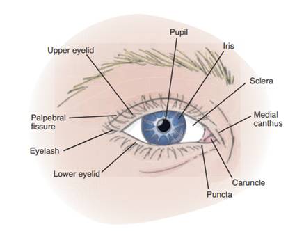
External structure of the eye
The eyelids join at two points: the lateral (outer) canthus and medial (inner) canthus. The medial canthus contains the puncta, two small openings that allow drainage of tears into the lacrimal system, and the caruncle, a small, fleshy mass that contains sebaceous glands. The white space between open eyelids is called the palpebral fissure. When closed, the eyelids should touch. When open, the upper lid position should be between the upper margin of the iris and the upper margin of the pupil. The lower lid should rest on the lower border of the iris. No sclera should be seen above or below the limbus (the point where the sclera meets the cornea).
Eyelashes are projections of stiff hair curving outward along the margins of the eyelids that filter dust and dirt from air entering the eye.
The conjunctiva is a thin, transparent, continuous membrane that is divided into two portions: a palpebral and a bulbar portion. The palpebral conjunctiva lines the inside of the eyelids, and the bulbar conjunctiva covers most of the anterior eye, merging with the cornea at the limbus. The point at which the palpebral and bulbar conjunctivae meet creates a folded recess that allows movement of the eyeball. This transparent membrane allows for inspection of underlying tissue and serves to protect the eye from foreign bodies.
The lacrimal apparatus consists of glands and ducts that serve to lubricate the eye. The lacrimal gland, located in the upper outer corner of the orbital cavity just above the eye, produces tears. As the lid blinks, tears wash across the eye then drain into the puncta, which are visible on the upper and lower lids at the inner canthus. Tears empty into the lacrimal canals and are then channeled into the nasolacrimal sac through the nasolacrimal duct. They drain into the nasal meatus.
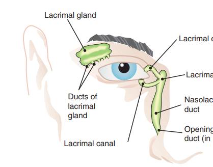
The lacrimal apparatus consists of tear glands and ducts
The extraocular muscles are the six muscles attached to the outer surface of each eyeball. These muscles control six different directions of eye movement. Four rectus muscles are responsible for straight movement, and two oblique muscles are responsible for diagonal movement. Each muscle coordinates with a muscle in the opposite eye. This allows for parallel movement of the eyes and thus the binocular vision characteristic of humans. Innervation for these muscles is supplied by three cranial nerves: the oculomotor (III) trochlear (IV), and abducens (VI).
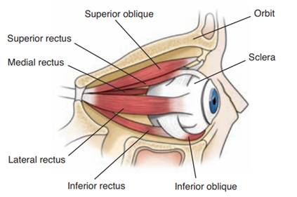
Extraocular muscles control the direction of eye movement
INTERNAL STRUCTURES OF THE EYE
The eyeball is composed of three separate coats or layers. The external layer consists of the sclera and cornea. The sclera is a dense, protective, white covering that physically supports the internal structures of the eye. It is continuous anteriorly with the transparent cornea (the “window of the eye”). The cornea permits the entrance of light, which passes through the lens to the retina. It is well supplied with nerve endings, making it responsive to pain and touch.
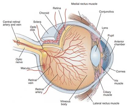
Anatomy of the eye
Because of this sensory property, contact with a wisp of cotton stimulates a blink in both eyes known as the corneal reflex. This reflex is supported by the trigeminal nerve, which carries the afferent sensation into the brain, and the facial nerve, which carries the efferent message that stimulates the blink.
The middle layer contains both an anterior portion, which includes the iris and the ciliary body, and a posterior layer, which includes the choroid. The ciliary body consists of muscle tissue that controls the thickness of the lens, which must be adapted to focus on objects near and far away.
The iris is a circular disc of muscle containing pigments that determine eye color. The central aperture of the iris is called the pupil. Muscles in the iris adjust to control the pupil’s size, which controls the amount of light entering the eye. The muscle fibers of the iris also decrease the size of the pupil to accommodate for near vision and dilate the pupil when far vision is needed.
The lens is a biconvex, transparent, avascular, encapsulated structure located immediately posterior to the iris. Suspensory ligaments attached to the ciliary body support the position of the lens. The lens functions to refract (bend) light rays onto the retina. Adjustments must be made in refraction depending on the distance of the object being viewed. Refractive ability of the lens can be changed by a change in shape of the lens (which is controlled by the ciliary body). The lens bulges to focus on close objects and flattens to focus on far objects.
The chorioid layer contains the vascularity necessary to provide nourishment to the inner aspect of the eye and prevents light from reflecting internally. Anteriorly, it is continuous with the ciliary body and the iris.
The innermost layer, the retina, extends only to the ciliary body anteriorly. It receives visual stimuli and sends it to the brain. The retina consists of numerous layers of nerve cells, including the cells commonly called rods and cones. These specialized nerve cells are often referred to as “photoreceptors” because they are responsive to light. The rods are highly sensitive to light, regulate black and white vision, and function in dim light. The cones function in bright light and are sensitive to color.
The optic disc is a cream-colored, circular area located on the retina toward the medial or nasal side of the eye. It is where the optic nerve enters the eyeball. The optic disc can be seen with the use of an ophthalmoscope and is normally round or oval in shape, with distinct margins. A smaller circular area that appears slightly depressed is referred to as the physiologic cup. This area is approximately one-third the size of the entire optic disc and appears somewhat lighter/whiter than the disc borders.
The retinal vessels can be readily viewed with the aid of an ophthalmoscope. Four sets of arterioles and venules travel through the optic disc, bifurcate, and extend to the periphery of the fundus. Vessels are dark red and grow progressively narrower as they extend out to the peripheral areas. Arterioles carry oxygenated blood and appear brighter red and narrower than the veins. The general background, or fundus, varies in color, depending on skin color. A retinal depression known as the fovea centralis is located adjacent to the optic disc in the temporal section of the fundus. This area is surrounded by the macula, which appears darker than the rest of the fundus. The fovea centralis and macular area are highly concentrated with cones and form the area of highest visual resolution and color vision.
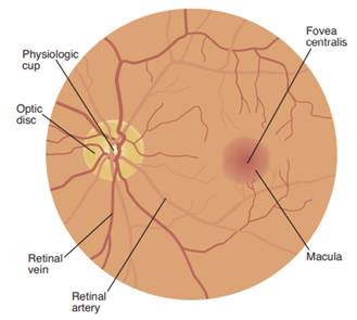
Normal ocular fundus.
The eyeball contains several chambers that serve to maintain structure, protect against injury, and transmit light rays. The anterior chamber is located between the cornea and iris, and the posterior chamber is the area between the iris and the lens. These chambers are filled with aqueous humor, a clear liquid substance produced by the ciliary body. Aqueous humor helps to cleanse and nourish the cornea and lens as well as maintain intraocular pressure. The aqueous humor filters out of the eye from the posterior to the anterior chamber then into the canal of Schlemm through a filtering site called the trabecular meshwork. Another chamber, the vitreous chamber, is located in the area behind the lens to the retina. It is the largest of the chambers and is filled with a vitreous humor that’s clear and gelatinous.
WORKING OF THE EYE
Light rays enter the eye through the cornea, the clear front “window” of the eye. The cornea’s refractive power bends the light rays in such a way that they pass freely through the pupil the opening in the center of the iris through which light enters the eye.
The iris works like a shutter in a camera. It has the ability to enlarge and shrink, depending on how much light is entering the eye.
After passing through the iris, the light rays pass thru the eye’s natural crystalline lens. This clear, flexible structure works like the lens in a camera, shortening and lengthening its width in order to focus light rays properly.
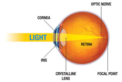
Light rays pass through a dense, transparent gel-like substance, called the vitreous that fills the globe of the eyeball and helps the eye hold its spherical shape.
In a normal eye, the light rays come to a sharp focusing point on the retina. The retina functions much like the film in a camera. It is responsible for capturing all of the light rays, processing them into light impulses through millions of tiny nerve endings, then sending these light impulses through over a million nerve fibers to the optic nerve.
Because the keratoconus cornea is irregular and cone shaped, light rays enter the eye at different angles, and do not focus on one point the retina, but on many different points causing a blurred, distorted image.
In summary, the cornea is the clear, transparent front covering which admits light and begins the refractive process. It also keeps foreign particles from entering the eye.
The pupil is an adjustable opening that controls the intensity of light permitted to strike the lens. The lens focuses light through the vitreous humor, a clear gel-like substance that fills the back of the eye and supports the retina.
The retina receives the image that the cornea focuses through the eye’s internal lens and transforms this image into electrical impulses that are carried by the optic nerve to the brain. We can tolerate very large scars on our bodies with no concern except for our vanity. This is not so in the cornea. Even a minor scar or irregularity in the shape can impair vision. No matter how well the rest of the eye is functioning, if the cornea is scarred, clouded or distorted, vision will be affected.
In keratoconus, the irregular shape of the cornea does not allow it to do its job correctly, leading to distortion of the image it passed to the retina and transmitted to the brain.
THE CORNEA
The eye is enclosed by a tough white sac, the sclera. The cornea is the transparent window in this white sac which allows the objects you are looking at to be carried in the form of light waves into the interior of the eye.
The surface of the cornea is where light begins its journey into the eye. The cornea’s mission is to gather and focus visual images. Because it is out front, like the windshield of an automobile, it is subject to considerable abuse from the outside world.
The cornea is masterfully engineered so that only the most expensive manmade lenses can match its precision. The smoothness and shape of the cornea, as well as its transparency, is vitally important to the proper functioning of the eye. If either the surface smoothness or the clarity of the cornea suffers, vision will be disrupted.
CORNEAL LAYERS
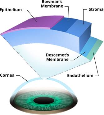
Although appearing to be one clear membrane, the cornea is composed of five distinct layers of tissue, each with its own function.
· Epithelium is the thin outermost layer of fast-growing and easily-regenerated cells.
· Bowman’s layer consists of irregularly-arranged collagen fibers and protects the corneal stroma. It is 8 to 14 microns thick.
· Stroma, the transparent middle and thickest layer of the cornea is made up of regularly-arranged collagen fibers and keratocytes (specialized cells that secrete the collagen and proteoglycans needed to maintain the clarity and curvature of the cornea)
· Descemet’s membrane is a thin layer that serves as the modified basement membrane of the corneal endothelium.
· Endothelium is a single layer of cells responsible for maintaining proper fluid balance between the aqueous and corneal stromal compartments keeping the cornea transparent.
VISION
Visual Fields and Visual Pathways
A visual field refers to what a person sees with one eye. The visual field of each eye can be divided into four quadrants: upper temporal, lower temporal, upper nasal, and lower nasal. The temporal quadrants of each visual field extend farther than the nasal quadrants. Thus, each eye sees a slightly different view but their visual fields overlap quite a bit. As a result of this, humans have binocular vision (“two eyed” vision) in which the visual cortex fuses the two slightly different images and provides depth perception or three dimensional vision.
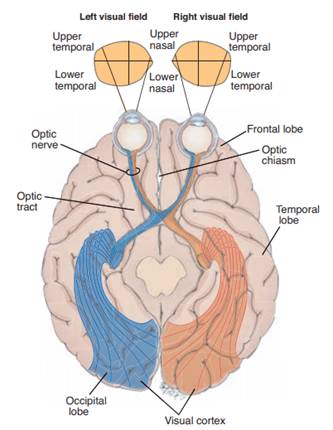
Visual fields and visual pathways. Each eye has a slightly different view of the same field. However, the views overlap significantly, which accounts for binocular vision.
Visual perception occurs as light rays strike the retina, where they are transformed into nerve impulses, conducted to the brain through the optic nerve, and interpreted. In the eye, light must pass through transparent media (cornea, aqueous humor, lens, and vitreous body) before reaching the retina.
The cornea and lens are the main eye components that refract (bend) light rays on the retina. The image projected on the retina is upside down and reversed right to left from the actual image. For example, an image from the lower temporal visual field strikes the upper temporal quadrant of the retina. At the point where the optic nerves from each eyeball cross—the optic chiasma—the nerve fibers from the nasal quadrant of each retina (from both temporal visual fields) cross over to the opposite side. At this point, the right optic tract contains only nerve fibers from the right side of the retina and the left optic tract contains only nerve fibers from the left side of the retina. Therefore, the left side of the brain views the right side of the world.
Visual Reflexes
The pupillary light reflex causes pupils immediately to constrict when exposed to bright light. This can be seen as a direct reflex, in which constriction occurs in the eye exposed to the light, or as an indirect or consensual reflex, in which exposure to light in one eye results in constriction of the pupil in the opposite eye. These protective reflexes, mediated by the oculomotor nerve, prevent damage to the delicate photoreceptors by excessive light.
Accommodation is a functional reflex allowing the eyes to focus on near objects. This is accomplished through movement of the ciliary muscles causing an increase in the curvature of the lens. This change in shape of the lens is not visible. However, convergence of the eyes and constriction of the pupils occur simultaneously and can be seen.
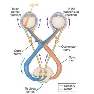
The pupils admit light that travels over the visual pathways. If a light focuses on only one eye, the pupil responds to ensure that the light needed for vision can enter but not so much that eye damage would result. The other pupil responds in the same manner. This phenomenon of direct pupillary response and consensual pupillary response is a reflex governed by the oculomotor nerve.
TYPES OF BLINKS
There are three types of blink.
Spontaneous blink
Spontaneous blinking which is done without external stimuli and internal effort. This type of blinking is conducted in the pre-motor brain stem and happens without conscious effort, like breathing and digestion. Blinking helps to spread the tear to all parts of our eye and helps to keep it moist. Our eyes should normally blink 15 times per minute.
Reflex blink
A reflex blink occurs in response to an external stimulus, such as contact with the cornea or objects that appear rapidly in front of the eye. A reflex blink is not necessarily a conscious blink either; however it does happen faster than a spontaneous blink.Reflex blink may occur in response to tactile stimuli (e.g. corneal, eyelash, skin of eyelid, contact with eyebrow), optical stimuli (e.g. dazzle reflex,or menace reflex) or auditory stimuli (e.g., menace reflex)
Reflex blinking may be caused by practically any peripheral stimulus, but the two functionally significant reflexes are (1) that resulting from stimulation of the endings of the fifth cranial nerve in the cornea, lid, or conjunctiva—the sensory blink reflex, or corneal reflex—and (2) that caused by bright light—the optical blink reflex. The corneal reflex is rapid (0.1 second reflex time) and is the last to disappear in deepening anesthesia, impulses being relayed from the nucleus of the fifth nerve to the seventh cranial nerve, which transmits the motor impulses. The reflex is said to be under the control of a medullary centre. The optical reflex is slower; in humans, the nervous pathway includes the visual cortex (the outer substance of the brain; the visual centre is located in the occipital—rear—lobe).
Voluntary blink
Voluntary blink is larger amplitude than Reflex blink, with the use of all 3 divisions of the orbicularis oculi muscle.