INTRODUCTION
Low vision is the loss of sight that is not correctible with prescription eyeglasses, contact lenses, or surgery. This type of vision loss does not include complete blindness, because there is still some sight and it can sometimes be improved with the use of visual aids.
Low vision includes different degrees of sight loss, from blind spots, poor night vision, and problems with glare to an almost complete loss of sight. The American Optometric Association defines low vision as two categories:
- "Partially sighted": the person has visual acuity between 20/70 and 20/200 with conventional prescription lenses.
- "Legally blind": the person has visual acuity no better than 20/200 with conventional correction and/or a restricted field of vision less than 20 degrees wide.
The ratio measurement of vision describes visual acuity, or the sharpness of vision, at 20 feet from an object. For example, having 20/70 vision means that you must be at 20 feet to see what a person with normal vision can see at 70 feet.
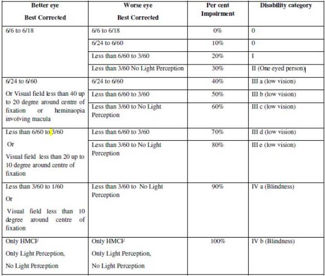
Low-vision means a condition where a person has any of the following conditions, namely:
1. visual acuity not exceeding 6/18 or less than 20/60 upto 3/60 or upto 10/200 (Snellen) in the better eye with best possible corrections; or
2. limitation of the field of vision subtending an angle of less than 40 degree up to 10 degree.
TYPES
Common Types of Low Vision
· Loss of Central Vision. The loss of central vision creates a blur or blind spot, but a person's side (peripheral) vision remains. This makes it difficult to read, recognize faces and distinguish most details in the distance. With side vision intact, however, mobility is usually unaffected.
· Loss of Peripheral (Side) Vision. People who lose their peripheral vision cannot distinguish anything to one side or both sides, or anything directly above and/or below eye level. Central vision remains, however, making it possible to see directly ahead, read and see faces. Typically, loss of peripheral vision affects mobility. If it is severe, it can slow reading speed because the person can only see a few words at a time. This is sometimes referred to as "tunnel vision."
· Blurred Vision. With blurred vision, both near and far vision is out of focus, even with the best possible correction with eyeglasses.
· Reduced Contrast Sensitivity. People with loss of contrast sensitivity, have a loss of vision quality. They tend to feel that there is a generalized haze with a sensation of a film or cloudiness.
· Glare Light Sensitivity. Occurs when standard levels of light overwhelm a person's visual system, producing a washed-out image and/or a glare. People with extreme light sensitivity may suffer pain or discomfort from relatively normal levels of light.
· Night Blindness. People with night blindness cannot see outside at night or in dimly lighted interior areas such as movie theaters or restaurants.
CAUSES OF LOW VISION
Eye diseases or conditions can cause visual impairment. Some of the more common causes of low vision include:
· Macular Degeneration. Macular degeneration is a disorder that affects the retina, the light-sensitive lining at the back of the eye where images are focused. The macula-the area on the retina responsible for sharp central vision-deteriorates, causing blurred vision. This can cause difficulty reading and, for some, a blurry or blind spot in the central area of vision.
· Cataracts. A cataract is a clouding of part or all the lens inside the eye. This clouding interferes with light reaching the retina at the back of the eye, resulting in general loss of vision. Causes include aging, long-term exposure to the sun's ultraviolet radiation, injury, disease and inherited disorders. If the eye is healthy, a cataract can be surgically removed. Usually, an intraocular lens implant is inserted in the eye, and vision is restored. Cataract surgery has a high success rate in otherwise healthy eyes. However, cataract surgery is not always possible for people who also have other eye diseases. These people may require low-vision rehabilitation to maximize their remaining vision.
· Glaucoma. Glaucoma causes damage to the optic nerve. Most commonly, this occurs due to increasing internal pressure in the eye because of problems with the flow or drainage of fluid within the eye. It can also occur when the internal pressure of the eye does not increase (normal-tension glaucoma), but there is not enough blood flow to the optic nerve. There are no early symptoms in the most common form of glaucoma, but the first signs of damage are defects in side (peripheral) vision and difficulty with night vision. If diagnosed early, it can be treated with drugs, or sometimes surgery can minimize vision loss.
· Diabetic Retinopathy. People with diabetes can experience day-to-day changes in their vision and/or visual functioning because of the disease. Diabetes can cause blood vessels that nourish the retina to develop tiny, abnormal branches that leak. This can interfere with vision and, over time, may severely damage the retina. Laser procedures and surgical treatments can reduce its progression but regulating blood sugar is the most important step in treating diabetic retinopathy.
· Retinitis Pigmentosa. Retinitis pigmentosa gradually destroys night vision, severely reduces side vision and may result in total vision impairment. An inherited disease, its first symptom-night blindness-usually occurs in childhood or adolescence.
· Amblyopia. In amblyopia, the visual system fails to develop normally during childhood. The blurry vision that results in one or both eyes is not easily corrected with normal glasses or contact lenses alone.
· Retinopathy of Prematurity (ROP). Retinopathy of prematurity occurs in infants born prematurely. It is caused by the high oxygen levels in incubators during the critical neonatal period.
· Retinal Detachment. With a retinal detachment, the retina separates from its underlying layer. It can cause total vision impairment in the affected eye. Causes include holes in the retina, eye trauma, infection, blood vessel disturbance or a tumor. If diagnosed early, most detached retinas can be surgically reattached with vision partially or completely restored.
· Acquired (Traumatic) Brain Injury. Vision can also be lost or damaged as a result of head injuries, brain damage and stroke. Signs and symptoms can include reduced visual acuity or visual field, contrast sensitivity, blurred vision, eye misalignment, poor judgment of depth, glare sensitivity, confusion when performing visual tasks, difficulty reading, double vision, headaches, dizziness, abnormal body posture and balance problems.
ASSESSMENT OF LOW VISION
Low vision is assessed only after taking all the possible measures to correct the vision as much as possible. These measures include medical and surgical interventions and/or use of spectacles/lenses.
CLASSIFICATION
The World
Health Organization uses the following classifications of visual
impairment.
When the vision in the better eye with best possible glasses correction is:
· 20/30 to 20/60, this is considered mild vision loss, or near-normal vision
· 20/70 to 20/160, this is considered moderate visual impairment, or moderate low vision
· 20/200 or worse, this is considered severe visual impairment, or severe low vision
· 20/500 to 20/1000, this is considered profound visual impairment or profound low vision
· Less than 20/1000, this is considered near-total visual impairment or near total low vision
No light perception, this is considered total visual impairment, or total blindness
DISABILITY CERTIFICATE FOR LOW-VISION
In India, under RPWD Act 2016, low vision is considered to be a disability. A person having benchmark disability, can avail disability benefits from the government.
If you think you have low vision and you want to get a disability certificate, you should visit a nearby government hospital for further directions.
The medical authority will decide whether disability certificate should be temporary or permanent. The disability shall be permanent to be certified. The certificate can be temporary if condition is likely to worsen and also for specific purposes such as for pursuing education. The need of reassessment, if required, should be clearly mentioned in the certificate with time frame. In certain cases such as keratoconus, developmental defects, operated congenital cataract with corneal decompensation, operated congenital glaucoma with hazy cornea etc., the patient especially can be issued a temporary certificate.
Medical Authority for Disability Certificate
The medical authority shall comprise of one ophthalmologist and certificate of disability shall be countersigned by Medical Superintendent or Chief Medical Officer or Civil Surgeon or any other equivalent authority as notified the State Government.
VISION CHARTS
Snellen Chart
Used to test distant visual acuity, the Snellen chart consists of lines of different letters stacked one on top of the other. The letters are large at the top and decrease in size from top to bottom. The chart is placed on a wall or door at eye level in a well-lighted area. The client stands 20 feet from the chart and covers one eye with an opaque card (which prevents the client from peeking through the fingers). Then the client reads each line of letters until he or she can no longer distinguish them.
E Chart
If the client cannot read or has a handicap that prevents verbal communication, the E chart is used. The E chart is configured just like the Snellen chart but the characters on it are only Es, which face in all directions. The client is asked to indicate by pointing which way the open side of the E faces. If the client wears glasses, they should be left on, unless they are reading glasses (reading glasses blur distance vision).
Test Results
Acuity results are recorded somewhat like blood pressure readings—in a manner that resembles a fraction (but in no way is interpreted as a fraction). A common example of an acuity test score is 20/20. The top, or first, number is always 20, indicating the distance from the client to the chart. The bottom, or second, number refers to the last full line the client could read. Usually the last line on the chart is the 20/20 line. The examiner needs to document whether the client wore glasses during the test. If any letters on a line are missed, encourage the client to continue reading until he or she cannot distinguish any letters, but record the number of letters missed by using a minus sign. If the client missed two letters on the 20/30 line, the recorded score would be 20/30 -2.
