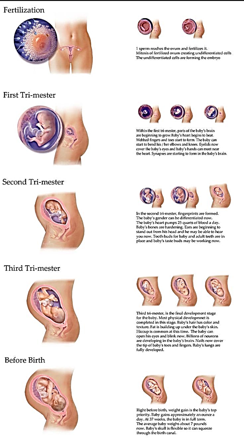
PRENATAL DEVELOPMENT
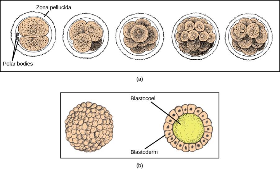
Fertilization is the process in which gametes (an egg and sperm) fuse to form a zygote. To ensure that the offspring has only one complete diploid set of chromosomes, only one sperm must fuse with one egg. In mammals, a layer called the zona pellucida protects the egg. At the tip of the head of a sperm cell is a structure like a lysosome called the acrosome, which contains enzymes. When a sperm binds to the zona pellucida, a series of events, called the acrosomal reactions, take place. These reactions, involving enzymes from the acrosome, allow the sperm plasma membrane to fuse with the egg plasma membrane and permit the sperm nucleus to transfer into the ovum. The nuclear membranes of the egg and sperm break down and the two haploid nuclei fuse to form a diploid nucleus or genome.
To ensure that no more than one sperm fertilizes the egg, once the acrosomal reactions take place at one location of the egg membrane, the egg releases proteins in other locations to prevent other sperm from fusing with the egg.
The development of multi-cellular organisms begins from this single-celled zygote, which undergoes rapid cell division, called cleavage, to form a hollow ball of cells called a blastula.
The Ovum
Conception occurs when an egg from the mother is fertilized by a sperm from the father. In humans, the conception process begins with ovulation, when an ovum, or egg (the largest cell in the human body), which has been stored in one of the mother’s two ovaries, matures and is released into the fallopian tube. Ovulation occurs about halfway through the woman‘s menstrual cycle and is aided by the release of a complex combination of hormones. In addition to helping the egg mature, the hormones also cause the lining of the uterus to grow thicker and more suitable for implantation of a fertilized egg.
The Zygote
Within several hours, half of the 23 chromosomes from the egg and half of the 23 chromosomes from the sperm fuse together, creating a zygote—a fertilized ovum. The zygote continues to travel down the fallopian tube to the uterus. Although the uterus is only about 4 inches away in the woman‘s body, this is nevertheless a substantial journey for a microscopic organism, and fewer than half of zygotes survive beyond this earliest stage of life. If the zygote is still viable when it completes the journey, it will attach itself to the wall of the uterus, but if it is not, it will be flushed out in the woman‘s menstrual flow. During this time, the cells in the zygote continue to divide: The original two cells become four, those four become eight, and so on, until there are thousands (and eventually trillions) of cells. Soon the cells begin to differentiate, each taking on a separate function. The earliest differentiation is between the cells on the inside of the zygote, which will begin to form the developing human being, and the cells on the outside, which will form the protective environment that will provide support for the new life throughout the pregnancy.
The Embryo
Once the zygote attaches to the wall of the uterus, it is known as the embryo. During the embryonic phase, which will last for the next 6 weeks, the major internal and external organs are formed, each beginning at the microscopic level, with only a few cells. The changes in the embryo‘s appearance will continue rapidly from this point until birth. While the inner layer of embryonic cells is busy forming the embryo itself, the outer layer is forming the surrounding protective environment that will help the embryo survive the pregnancy. This environment consists of three major structures: The amniotic sac is the fluid-filled reservoir in which the embryo (soon to be known as a fetus) will live until birth, and which acts as both a cushion against outside pressure and as a temperature regulator. Theplacenta is an organ that allows the exchange of nutrients between the embryo and the mother, while at the same time filtering out harmful material. The filtering occurs through a thin membrane that separates the mother‘s blood from the blood of the fetus, allowing them to share only the material that is able to pass through the filter. Finally, the umbilical cord links the embryo directly to the placenta and transfers all material to the fetus. Thus the placenta and the umbilical cord protect the fetus from many foreign agents in the mother‘s system that might otherwise pose a threat.
The Fetus
Beginning in the 9th week after conception, the embryo becomes a fetus. The defining characteristic of the fetal stage is growth. All the major aspects of the growing organism have been formed in the embryonic phase, and now the fetus has approximately six months to go from weighing less than an ounce to weighing an average of 6 to 8 pounds. That‘s quite a growth spurt.
The fetus begins to take on many of the characteristics of a human being, including moving (by the 3rd month the fetus is able to curl and open its fingers, form fists, and wiggle its toes), sleeping, as well as early forms of swallowing and breathing. The fetus begins to develop its senses, becoming able to distinguish tastes and respond to sounds. Research has found that the fetus even develops some initial preferences. A newborn prefers the mother‘s voice to that of a stranger, the languages heard in the womb over other languages (DeCasper & Fifer, 1980; Moon,)